Chapter 8. Measuring Velocity Using the CAM1
- Table of Contents
- Selecting a Capillary
- Taking Measurements
Selecting a Capillary
First the laser position will need to be marked on the video monitor. Focus onto the surface of the skin before applying any oil. The laser beam should be just visible as a small bright spot in the centre of the video image. It may be necessary to turn down the light source. If you have the CapiScope option mark the laser spot with the crosshair, otherwise mark circle on the video monitor using a dry marker pen.
Position a perpendicular capillary loop under the laser beam. Choose a capillary where there is little surface reflection of the laser. Too much surface reflection will feedback into the laser causing a lot of 'noise'. This sounds like loud crackling. Too much surface reflection may also cause tissue surface movements to dominate the detected signal. Try adding more optical matching oil, or slightly adjust the angle of the skin tissue. It may still be possible to acquire good measurements but it is difficult and tiring if using the audio output to position the beam. When the beam is moved off the capillary a zero or close to zero flow should be shown with little noise.
Note that it is quite common for there to be little or no capillary flow when the room is cool or the subject is nervous. In this case there will be no difference when the laser beam is on or off position. Therefore when first using the CAM1, experiment with subjects with warm hands, or stimulate high velocities by pinching or scraping the surface. Faster flows also give a much stronger signal, so making them easier to practice with. Slow velocities require that the subject is very still, otherwise it will be difficult to distinguish between capillary flow and tissue movement.
Position over the top of the arterial or venous limb. For low flows this may be determined by noting the direction of flow on the video monitor. For higher flows or skin with poor images this may not be possible. One should then either measure both limbs, or allow for a lower overall average reading assuming that half of measurements may be from the arterial limb and half from the venous limb.
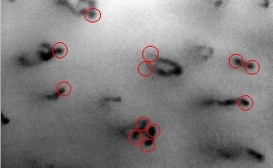
This image shows some of the possible sites which should give good Doppler signals. Note the capillary in the centre of the image. This capillary loop folds over near the surface, but the arterial and venous limbs should give good signals. Even though they cannot be visualised in the image because of scattering in the tissue, the longer wavelength laser is scattered less and enough signal can sometimes be obtained to give good measurement.
The four measurement points in the bottom centre should be treated with care. Since the two capillaries are so close together, some signal may be picked up from the adjacent vessel. This should be visible in the Doppler spectrum as two peaks in the spectrum.
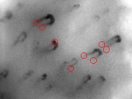
In this image, taken close to the nailfold, capillaries are folding over at the tops of the loops. Here the laser should be positioned at the point where the capillaries start to bend.
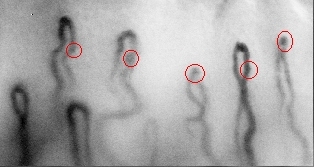
At the nailfold, the flow is perpendicular to the laser beam, so there will be no Doppler shift. However, light may be reflected along the axis of the capillary and be shifted. Try the apex of capillaries which are twisted sideways, or points on tortuous loops which travel perpendicular to the surface and show up as darker points.